
TAGC 2020
TAGC 2020
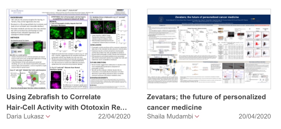
Daria Lukasz (1, 2,) Katie S. Kindt (2)
1) National Institutes of Health – Johns Hopkins University Graduate Partnerships Program, NIH, Bethesda, MD; 2) National Institute on Deafness and Other Communication Disorders, NIH, Bethesda, MD.
The powerful gram-negative aminoglycoside antibiotic neomycin specifically targets not only life-threatening bacterial infections but also sensory hair cells. Hair-cell death causes irreversible and often isolating hearing loss in humans. Though much is known about the process of neomycin-induced cell death, factors that underlie hair-cell neomycin susceptibility have not been fully elucidated. Current work suggests that persistent metabolic activity resulting from the near constant stimulation of apical mechanotransduction channels in an aqueous environment may sensitize cells to neomycin by contributing to a slow buildup of toxic metabolic byproducts. Additionally, mechanosensation has been shown to stress mitochondria and subsequently boost the production of reactive oxygen species (ROS). Neomycin itself triggers ROS overproduction because it targets mitochondria and thus may pose a direct threat to metabolically stressed cells. Our work aims to understand the relationship between hair-cell metabolic activity, ROS production, and neomycin susceptibility.
To explore this further, we utilized a larval zebrafish model which offers external lateral-line hair cells that express membrane-localized GCaMP6s and allows for close monitoring of individual cells in an in vivo and in toto context. Hair cells rely on the influx of calcium through basal voltage-gated Cav1.3 channels for signal transduction, and so we began by acutely blocking these channels with an antagonist isradipine during neomycin bath treatment. We found that acute calcium block did not prevent cell death relative to control. However, examination of cav1.3-/- mutants revealed augmented cell survival, suggesting that the chronic block of metabolic activity does mitigate neomycin susceptibility. Preliminary data utilizing the cytosolic ROS indicator cellROX Orange also shows a reduction in oxidative stress in these mutants. Furthermore, examination of otof-/- mutants, which lack the calcium sensor necessary for the coupling of calcium influx with vesicle fusion, revealed that the loss of exocytosis alone reduces neomycin susceptibility. These findings suggest that metabolic activity related to the exocytosis of glutamate-containing vesicles contributes to oxidative stress that weakens hair cells. We are currently examining other activity-related mutations and more specific ROS indicators with the ultimate goal of identifying an otoprotective therapeutic target and preventing hearing loss.
Shaila Mudambi (1), Jessica Miller (2), Saumya Kasliwal(1), Seray Er(1), Kamden Gray(1), Ceylan Metin(1), Russell Sillmon(1), Anna Zdunek(1), Mary Pasquale(1), Michael Pishvaian(1), Stephen Byers(1), Eric Glasgow(1)
1) Georgetown University; 2) Vanderbilt University Medical Center.
Personalized medicine offers a more informed strategy to treat cancer patients. Although promising, the efficacy of targeting therapy based on a patient’s molecular profile is still difficult to predict. Currently, ex vivo models such as patient derived mouse xenograft (mPDX), organoid culture, and conditionally reprogrammed cells (CRC) are being developed to improve the prediction of chemotherapy response. These models require significant expansion of tumor cells for drug testing requiring relatively long wait times, which introduces potential for changes in genetic and epigenetic characteristics of the tumor. Moreover, these models do not consider the tumor microenvironment, including the immune system which is important for cancer therapy. Therefore, to compliment these models, we have developed patient-derived xenografts in zebrafish embryos or ‘Zevatars’ that directly samples the patient’s tumor, is rapid enough to benefit patients with aggressive disease, allows for high-throughput drug screening, and provides a readout that is not simply a surrogate for cell growth. The assay is performed by implanting labelled pieces of patient tumor tissue (fresh or cryopreserved) into 2 dpf embryos, treating with drugs, monitoring tumor behaviors, including size changes, cell migration, and metastasis through imaging. The best treatment for a given patient can be determined in less than one week. In addition, we can generate Zevatars from multiple tumor types including pancreatic cancer and liver metastasis, and are expanding our repertoire to breast, lung, ovarian and squamous epithelial cancers. Our results show that tumor biopsy tissue differentially responds to standard of care drugs. Importantly, the response varies greatly from patient to patient, thus recapitulating patient tumor behavior in the clinic. However, one major limitation of the Zevatar is that we cannot study adaptive immune responses or cancer immunotherapy in this model, though we have observed macrophage homing to cancer xenografts suggesting a role for the innate immune system. Hence we are developing a humanized fish in which we replace the zebrafish hematopoietic system with a human hematopoietic system via stem and progenitor cell transplantation (HSPC). If successful we can then predict chemotherapy and immunotherapy effectiveness by using a Zevatar with a patient’s own HSPCs coupled with their tumor biopsy. In developing this technology we can provide a rapid and relatively cost-effective method for personalized cancer diagnostics.
Sponsor Message

Join our community of nearly 6,000 researchers from all career stages and more than 50 countries. You’ll receive discounted conference fees and become eligible for travel awards and professional development programs.